G. Zhang, S. Harput, S. Lin, K. Christensen-Jeffries, C.H. Leow, J. Brown, C. Dunsby, R. Eckersley, M. Tang, “Acoustic Wave Sparsely-Activated Localization Microscopy (AWSALM): Super-Resolution Ultrasound Imaging using Acoustic Activation and Deactivation of Nanodroplets”
S. Harput, K. Christensen-Jeffries, J. Brown, Y. Li, K.J. Williams, A.H. Davies, R.J. Eckersley, C. Dunsby, and M-X. Tang, “Two-stage Motion Correction for Super-Resolution Ultrasound Imaging in Human Lower Limb”
Applied Physics Letter (vol. 113, no. 1)
Affiliations:
G. Zhang, S. Harput, S. Lin, C.H. Leow, M. Tang : ULIS Group, Department of Bioengineering, Imperial College London, London, SW7 2AZ, UK
K. Christensen-Jeffries, J. Brown, and R. Eckersley: Biomedical Engineering Department, Division of Imaging Sciences, King’s College London, SE1 7EH, London, UK
C. Dunsby: Department of Physics and the Centre for Pathology, Imperial College London, London, SW7 2AZ, UK
Abstract:
Photo-activated localization microscopy (P ALM) has revolutionized the field of fluorescence microscopy by breaking the diffraction limit in spatial resolution. In this study, ‘acoustic wave sparsely-activated localization microscopy (AWSALM)’, an acoustic counterpart of PALM, is developed to super-resolve structures which cannot be resolved by conventional B-mode imaging. AWSALM utilizes acoustic waves to sparsely and stochastically activate decafluorobutane nanodroplets by acoustic vaporization, and to simultaneously deactivate the existing vaporized nanodroplets via acoustic destruction. In this method, activation, imaging, and deactivation are all performed using acoustic waves. Experimental results show that sub-wavelength micro-structures not resolvable by standard B-mode ultrasound images can be separated by AWSALM. This technique is flow independent and does not require a low concentration of contrast agents, as is required by current ultrasound super resolution techniques. Acoustic activation and deactivation can be controlled by adjusting the acoustic pressure, which remains well within the FDA approved safety range. In conclusion, this study shows the promise of a flow and contrast agent concentration independent super-resolution ultrasound technique which has potential to be faster and go beyond vascular imaging.
July 2018 front cover image of the Applied Physics Letter
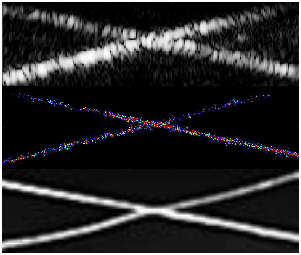
Figure shows (Top) B-Mode, (Middle) Super-resolution and (Bottom) optical images of activated decafluorobutane nanodroplets by acoustic vaporization in a cross section of 200µm tubes.
Supporting Bodies:
This work was supported in part by the EPSRC under Grant EP/N015487/1, EP/N014855/1 and EP/M011933/1 and in part by the Cancer Research UK Multidisciplinary Project Award C53470/A22353.