Background
In recent years, echo particle image velocimetry (ePIV), also known as ultrasound particle image Velocimetry (uPIV), has shown to be able to map flow velocity distribution in vivo. The technique provides valuable information about blood flow and flow induced stress on vessel wall for studying cardiovascular diseases. Using sequential ultrasound B-mode (or contrast specific) images, uPIV technique tracks either individual flow tracer (e.g. microbubble contrast agents) or speckle patterns within blood vessels in order to obtain local velocity vectors using cross-correlation, filtering, interpolation and other data analysis techniques.
Comparing to existing techniques such as optical PIV, Ultrasound Doppler or phase-contrast magnetic resonance imaging, uPIV has many advantages including: capable of imaging in turbid media (tissue), being angle independent and having high temporal resolution.
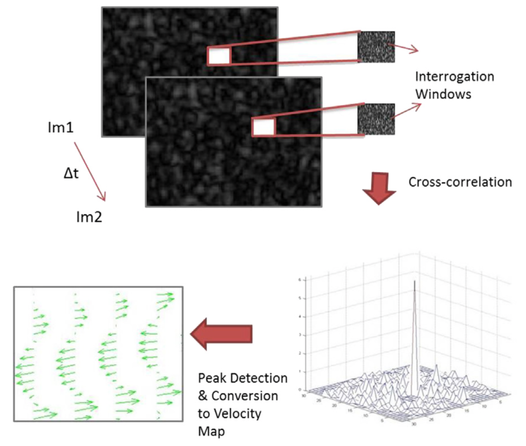
Figure 1 : Principle underlying conventional particle image velocimetry: Two consecutive ultrasound images were divided into several interrogation windows. For each window, cross-correlation analysis was performed to compute a local velocity displacement. The location of the peak within the correlation map was identified and displayed on the velocity map
Challenges
Although in vitro validation and some clinical applications of uPIV have been reported, the accuracy of the method to provide quantitative measurement is still limited due to e.g., the high dynamic range of the velocities to be measured, loss of information when acquiring in 2D for 3D structures, and for most existing clinical system, a relatively slow and line-by-line image acquisition.
Research
Investigation of errors in velocity estimation
In most existing clinical ultrasound imaging system an image is produced through line-by-line data acquisition. Such “beam sweeping” can introduce errors/biases to flow velocity estimation. We have investigated the significance of such errors under various flow and scanning conditions (Zhou et al. Ultrasound in Medicine and Biology, in press).
Next generation of uPIV – simulation and experiments
The state-of art of ultrasound imaging system to provide a high temporal resolution is being investigated. The quantitative measurement of conventional scanning ultrasound and some more advanced techniques such as Synthetic Aperture and Plane Wave Imaging are being investigated in both simulation and experiments to provide the highest possible accuracy, temporal and spatial resolution in PIV analysis. Both numerical and physical flow phantoms are developed for investigating the uPIV system.
In vivo application
Initial in vivo results
The developed techniques will be applied to in vivo applications in order to study various cardiovascular diseases. The following figure shows the initial in vivo results that have been developed in 2015 [1].
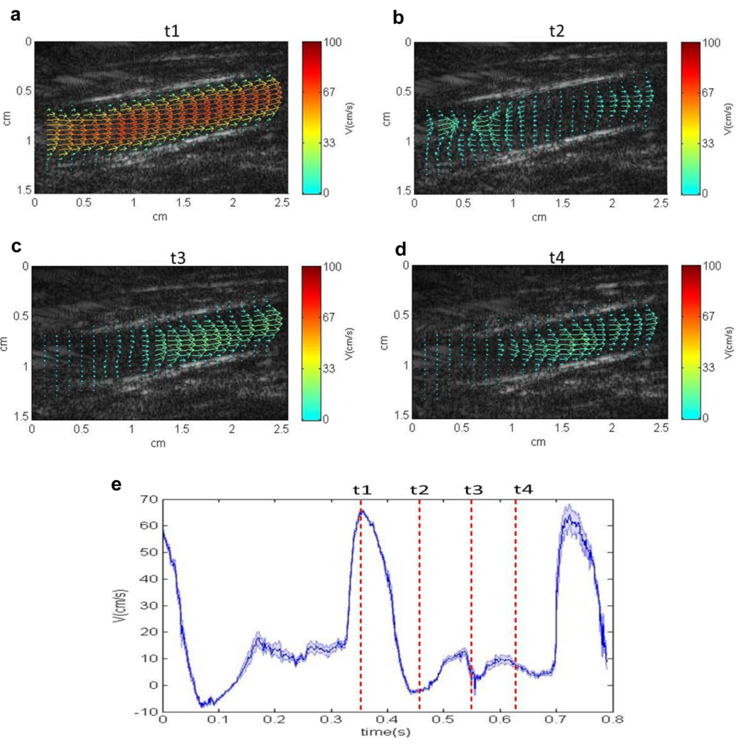
Figure 2 – (a–d) Quantitative visualisation of the flow patterns within a rabbit aorta. (e) Centreline velocity plot obtained using ultrasound imaging velocimetry from a 1 3 1-mm range gate within the aorta. The relative position of each flow pattern is marked in the velocity plot.
Recent in vivo results : flow imaging for atherosclerosis research
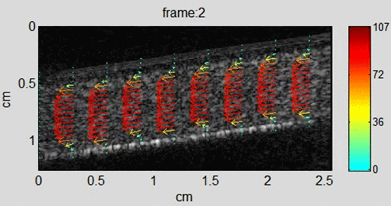
Atherosclerosis is a condition in which the blood vessels become narrowed or blocked due to the build-up of plaque, which is a mixture of fatty substances, cholesterol, and other materials. Wall shear stress (WSS) is the force exerted by blood flow on the inner surface of blood vessels. There is evidence to suggest that WSS plays a role in the development and progression of atherosclerosis. uPIV can not only provide detailed information about the flow patterns in the blood vessels, including the direction and velocity of blood flow but it can also be used to identify areas of low or high WSS. We have demonstrated the use of uPIV to measure wall shear stress accuratetly with and without the use of contrast agents both in 2D and in 3D [2, 3, 4].
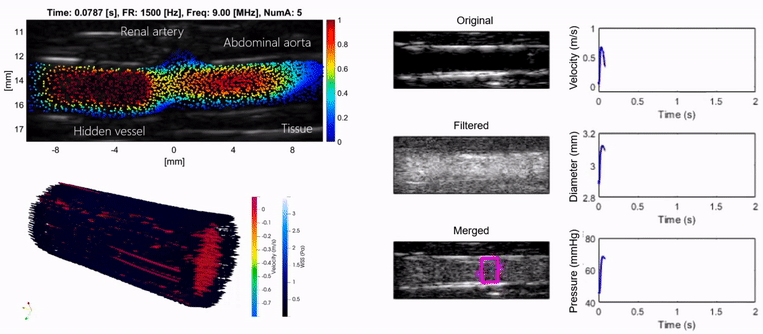
Figure 3 – Ultrasound Particle Image Velocimetry in the rabbit aorta in 2D, 3D, with and without contrast agents.
References
- Chee Hau Leow, Eleni Bazigou, Robert J. Eckersley, Alfred C.H. Yu, Peter D. Weinberg, Meng-Xing Tang, Flow Velocity Mapping Using Contrast Enhanced High-Frame-Rate Plane Wave Ultrasound and Image Tracking: Methods and Initial in Vitro and in Vivo Evaluation, Ultrasound in Medicine & Biology, Volume 41, Issue 11, November 2015, Pages 2913-2925
-
K Riemer, EM Rowland, CH Leow, MX Tang, PD Weinberg, Determining haemodynamic wall shear stress in the rabbit aorta in vivo using contrast-enhanced ultrasound image velocimetry, Annals of Biomedical Engineering 48 (6), 1728-1739
-
K Riemer, EM Rowland, J Broughton-Venner, CH Leow, M Tang, PD Weinberg, Contrast Agent-Free Assessment of Blood Flow and Wall Shear Stress in the Rabbit Aorta using Ultrasound Image Velocimetry, Ultrasound in Medicine & Biology 48 (3), 437-449
-
K Riemer, M Toulemonde, EM Rowland, CH Leow, MX Tang, PD Weinberg, 4D blood flow and wall shear stress measured using volumetric ultrasound image velocimetry, 2020 IEEE International Ultrasonics Symposium (IUS), 1-4