The development of microbubble contrast agents for ultrasound (US) imaging has made visualisation and quantification of blood flow in micro-vasculature possible. Such imaging technique can be applied to the diagnosis and treatment of a wide range of clinical conditions such as cardiovascular diseases and cancer.
Recently adapting the microbubbles to target specific molecules within the body has opened up a whole new area of molecular imaging using ultrasound and this is revolutionising the role that ultrasound imaging can play in biological and medical applications. Studies have shown that microbubbles can be made to target molecules associated with pathological/ physically conditions such as inflammation and angiogenesis (growth of vessels) and in vivo images visualising the distribution of target molecules has been initially demonstrated.
The extremely high sensitivity of ultrasound to microbubbles make ultrasound molecular imaging a highly attractive technique, which has the potential to detect diseases at its very early stage. The non-invasive, safe and cost effect properties of US imaging that have made this modality so successful and widespread for anatomical imaging are just as relevant in terms of molecular imaging.
Challenges / Opportunities
Currently ultrasound molecular imaging is still in its infancy and some challenges exist. Our understanding of the targeted microbubbles before, during and after they attach to their targets under the complex in vivo environment (e.g. being close to a vessel wall, under flow, and being driven by ultrasound etc) are still very limited. Such understanding will help the development of microbubbles with optimised targeting efficiency and development of detection and analysis strategies.
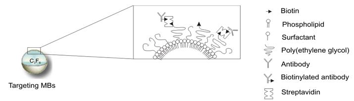
which rely on surface conjugation of monoclonal antibodies or other targeting ligands. PEG, polyethyleneglycol.
Research
Adherence of microbubbles under flow
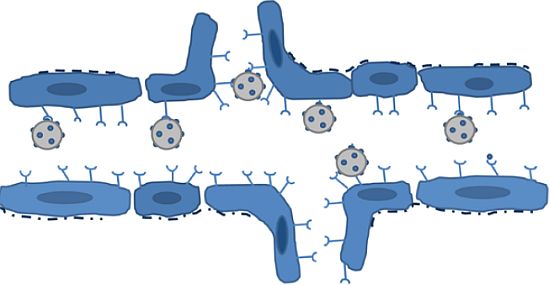
We have investigated the adherence of targeted microbubbles under flow conditions. The following is a movie showing the attachment of the targeted bubbles within a coated flow cell under microscope.
[youtube]https://www.youtube.com/watch?v=1Ap2rz4FfbE[/youtube]
Our results suggest that more targeted microbubbles adhere under pulsatile flow versus steady flow (Sennoga 2014 UMB).
Effects of ultrasound on adherent microbubbles
Ultrasound is used for detecting adherent microbubbles but does the ultrasound affect them?
[youtube]https://www.youtube.com/watch?v=DOiITv6YfMM[/youtube]
Our research shows that ultrasound can significantly affect adherent microbubbles by detaching them from their targets and / or deflating them. Loughran 2012 PMB
Detection of adherent microbubbles
We have investigated techniques to detect and distinguish attached bubbles from background (including tissue and free bubbles). In particular we have developed non-linear Doppler techniques which are capable of distinguishing both type of targets (tissue versus bubble) and motion of targets (in motion versus stationary/slow motion).
Mahue 2011 UFFC
Acoustic characterisation of adherent microbubbles
We have shown a difference in harmonic generation between microbubbles that are adherent versus those that are not. This could be used for detection of adherent microbubbles.
Casey 2013 UMB