M. Toulemonde, Y. Li, S. Lin, F. Cordonnier, M. Butler, W. C. Duncan, R. J. Eckersley, V. Sboros, and M-X. Tang, “High-Frame-Rate Contrast Echocardiography using diverging waves: initial in-vitro and in-vivo evaluation”
IEEE Transactions on Ultrasonics, Ferroelectrics, and Frequency Control, 2018
DOI: 10.1109/TUFFC.2018.2856756
Affiliations:
M. Toulemonde, Y. Li, S. Lin, F. Cordonnier, and M. X. Tang are with the ULIS Group, Department of Bioengineering, Imperial College London, London, SW7 2AZ, UK
R. J. Eckersley are with the Biomedical Engineering Department, Division of Imaging Sciences, King’s College London, SE1 7EH, London, UK
M. Buttler and V. Sboros, are with the Institute of Biological Chemistry Biophysics, and Bioengineering, Heriot-Watt University, Edinburgh, EH14 4AS
W C. Duncan is with the MRC Centre for Reproductive Health, The University of Edinburgh, Edinburgh EH16 4TJ
Abstract:
Contrast Echocardiography (CE) ultrasound with microbubble contrast agents (UCA) has significantly advanced our capability for assessment of cardiac function, including myocardium perfusion quantification. However in standard CE techniques obtained with line by line scanning, the frame rate and image quality are limited. Recent research has shown significant frame rate improvement in non-contrast cardiac imaging.
In this work we present and initially evaluate, both in-vitro and in-vivo, a high frame rate (HFR) CE imaging system using diverging waves and pulse inversion sequence. An imaging frame rate of 5500 frames per second before and 250 frames per second after compounding is achieved. A destruction-replenishment sequence has also been developed. The developed HFR CE is compared with standard CE in-vitro on a phantom and then in-vivo on a sheep heart.
The image signal to noise ratio, contrast between the myocardium and the chamber are evaluated. Results show up to 13.4 dB improvement in contrast for HFR CE over standard CE when compared at the same display frame-rate even when the average spatial acoustic pressure in HFR CE is 36% lower than the standard CE. It is also found that when coherent compounding is used the HFR CE image intensity can be significantly modulated by the flow motion in the chamber.
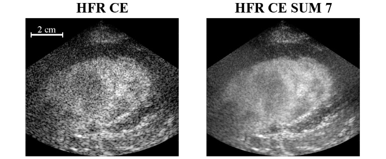
(Left) CE, (Middle) HFR CE and (Right) HFR CE SUM 7 videos of in-vivo heart sheep experiments. SUM 7 corresponds to the averaging of 7 envelope detected HFR CE frames, with a triangular window centered on the interested frame. CE and HFR CE are two successive acquisition at the same position. All videos are normalized and log compress by their own maximum and display with a dynamic range of 40 dB. The maximum depth is 90 mm. and the lateral axis cover 100 mm.
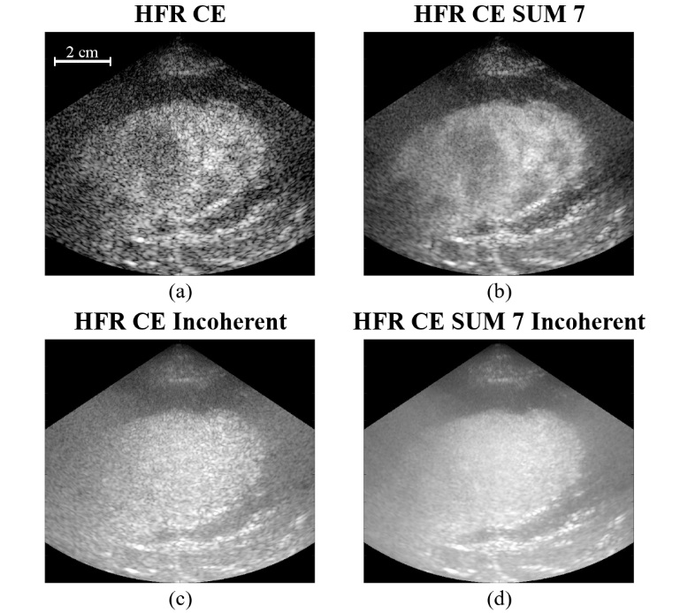
Figure shows HFR coherent (a, b) and incoherent (c, d) compounding and without and with temporal average of 7 consecutives frames in (a, c) and (b, d), respectively. All images are displayed with a 40 dB dynamic range. As expected, incoherent compounding removed the motion artefacts inside the chamber because phase information is lost, and so, small misalignment between echoes from each steered transmission does not significantly impact the image. However the image dynamic range and contrast are significant reduced, and some fine details inside the myocardium disappears.
Supporting Bodies:
This work was supported by an Engineering and Physical Sciences Research Council (EPSRC) grant EP/M011933/1 and grant EP/M010961/1.