Matthieu Toulemonde, Richard Corbett, Virginie Papadopoulou, Navtej Chahal, Yuanwei Li, Chee Hau Leow, David O. Cosgrove, Robert J. Eckersley, Neill Duncan, Roxy Senior, Meng-Xing Tang, “High Frame-Rate Contrast Echocardiography: In-Human Demonstration”
JACC: Cardiovascular Imaging
https://doi.org/10.1016/j.jcmg.2017.09.011
Matthieu Toulemonde, Yuanwei Li, Chee Hau Leow, Virginie Papadopoulou, Meng-Xing Tang
Department of Bioengineering, Imperial College London, London, SW7 2AZ UK
Richard Corbett, Neill Duncan
Hammersmith Hospital, Imperial College Healthcare NHS Trust London W12 0HS
David O. Cosgrove
Department of Medicine, Imperial College London, United Kingdom
Robert J. Eckersley
Biomedical Engineering Department of the Division of Imaging Sciences at King’s College London, London SE1 7EH
Navtej Chahal, Roxy Senior
Department of Echocardiography, Royal Brompton Hospital, London, SW3 6NP UK.
This work is funded by the UK Engineering and Physical Sciences Research Council grant EP/M011933/1 and grant EP/M010961/1 and the NIHR Imperial Biomedical Research Centre.
Keyword
High Frame Rate Ultrasound, Ultrafast, Microbubble Contrast Agents, CEUS, Echocardiography, Imaging, Flow Quantification
The video below shows the conventional focus CE acquisition versus the HFR CE method proposed. Focus-CE is acquired with a frame rate of 25 frames per seconds while HFR-CE is acquired at 250 frames per seconds.
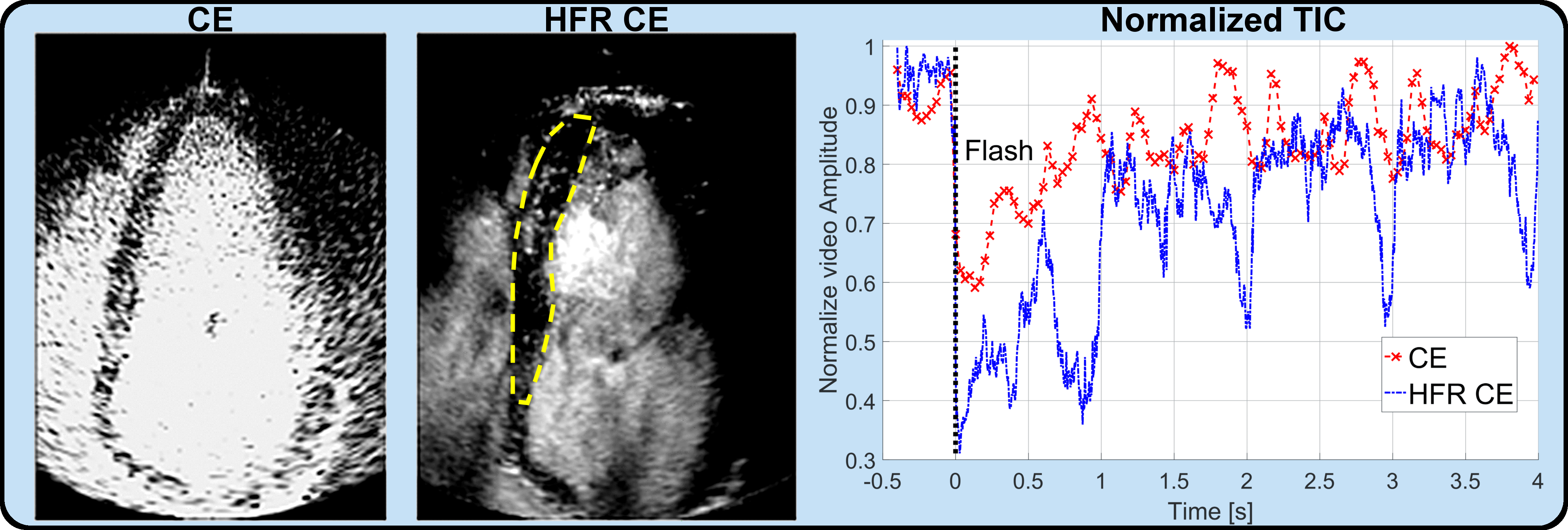
Figure 1: Replenishment time-intensity curves in healthy human volunteers
![]()
Figure 2: Chamber flow velocity map at different cardiac phases before and after R wave (R-0.12s, R-0.02s and R+0.12s respectively)