C. H. Leow, N. L. Bush, A. Stanziola, M. Braga, A. Shah, J. Hernández-Gil, N. J. Long, E. O. Aboagye, J. C. Bamber and M.X. Tang, “3D microvascular imaging using high frame rate ultrasound and ASAP without contrast agents: development and initial in vivo evaluation on
IEEE Transactions on Ultrasonics Ferroelectrics and Frequency Control PP(99):1-1
DOI: 10.1109/TUFFC.2019.2906434
Affiliation
C.H. Leow, A. Stanziola and M-X. Tang
N.L.Bush, A. Shah and J.C. Bamber are with the Joint Department of Physics and Cancer Research UK Cancer Imaging Centre, Institute of Cancer Research and Royal Marsden NHS Foundation Trust, United Kingdom.
M. Braga, J. Hernández-Gil, N. J. Long and E.O. Aboagye are with the Comprehensive Cancer Imaging Centre, Department of Surgery & Cancer, United Kingdom.
J. Hernández-Gil and N. J. Long are also with the Department of Chemistry, Imperial College London, United Kingdom.
Abstract:
Three-dimensional imaging is valuable to non-invasively assess angiogenesis given the complex 3D architecture of vascular networks. The emergence of high frame rate (HFR) ultrasound, which can produce thousands of images per second, has inspired novel signal processing techniques and their applications in structural and functional imaging of blood vessels. Although highly sensitive vascular mapping has been demonstrated using ultrafast Doppler, the detectability of microvasculature from the background noise may be hindered by the low signal to noise ratio (SNR) particularly in
Supporting Bodies:
This work is funded by the CRUK Multidisciplinary Project Award (C53470/A22353). C.H Leow would like to acknowledge NVIDIA Corporation for the donation of Titan XP GPU used in this research.
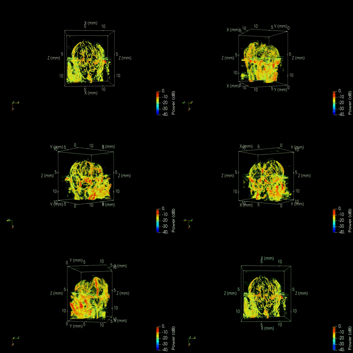
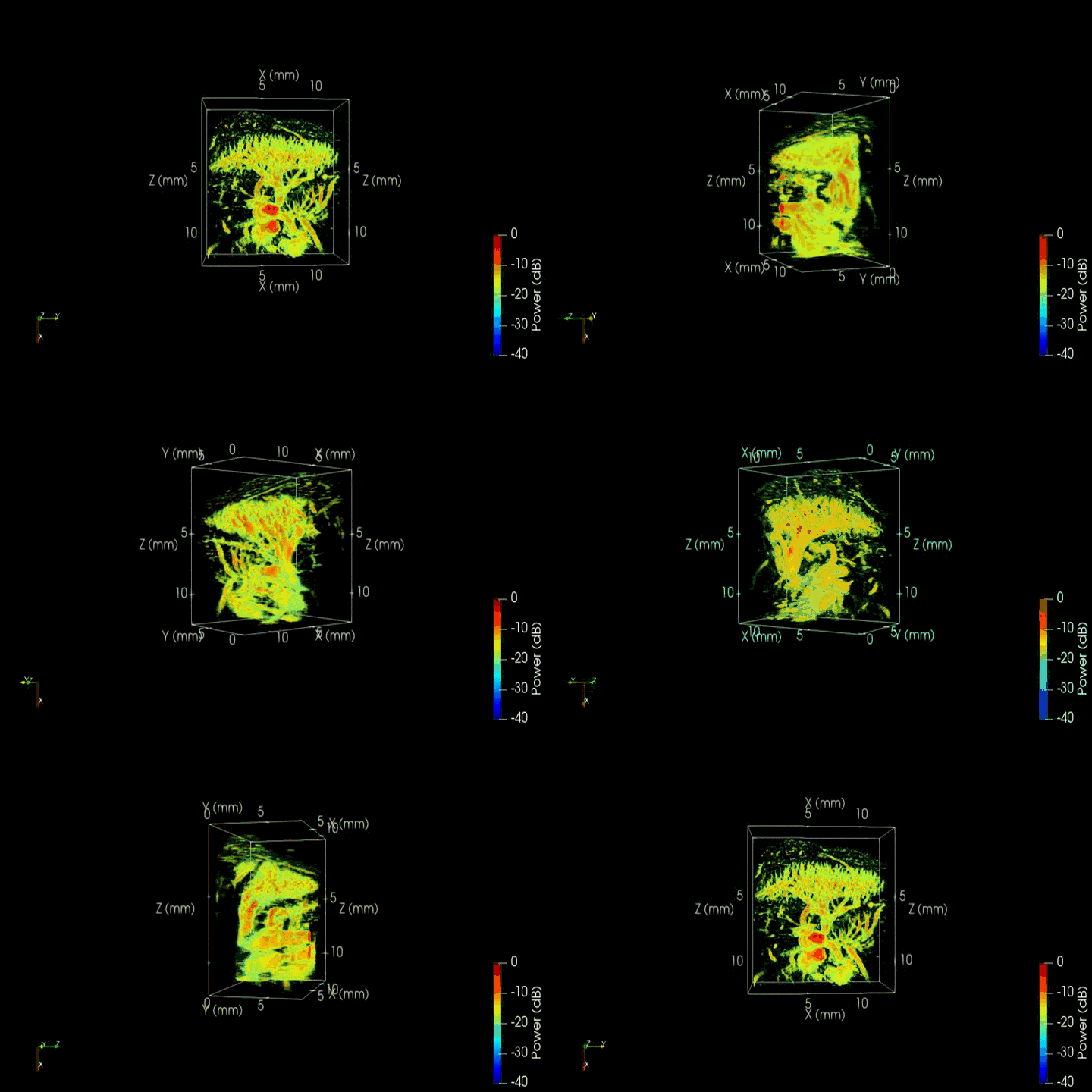
Fig: Volume render presentation of 3D microvascular images of a non-tumour kidney (left), liver (middle), and tumour (right) mouse model.


