X. Zhou, C.H. Leow, E. Rowland, K. Riemer, JM. Rubin, Peter D. Weinberg, M. Tang, “3D velocity and volume flow measurement in vivo using speckle decorrelation and 2D high frame rate contrast-enhanced ultrasound”
IEEE Transactions on Ultrasonics, Ferroelectrics, and Frequency Control, 2018
DOI: 10.1109/TUFFC.2018.2850535
Affiliations:
X Zhou, CH Leow, E Rowland, K Riemer, PD Weinberg and MX Tang are with the Department of Bioengineering, Imperial College London, London SW7 2AZ, UK
JM Rubin is with Department of Radiology, University of Michigan, Ann Arbor, MI 48109-5667, USA
Abstract:
Being able to measure 3D flow velocity and volumetric flow rate effectively in the cardiovascular system is valuable but remains a significant challenge in both clinical practice and research. Currently there has not been an effective and practical solution to the measurement of volume flow using ultrasound imaging systems due to challenges in existing 3D imaging techniques and high system cost.
In this study, a new technique for quantifying volumetric flow rate from the cross-sectional imaging plane of the blood vessel was developed by using speckle decorrelation, 2D high frame rate imaging with a standard 1D array transducer, microbubble contrast agents, and ultrasound imaging velocimetry (UIV). Through speckle decorrelation analysis of microbubble signals acquired with a very high frame rate and by using UIV to estimate the two in-plane flow velocity components, the third and out-of-plane velocity component can be obtained over time and integrated to estimate volume flow. The proposed technique was evaluated on a wall-less flow phantom in both steady and pulsatile flow. UIV in the longitudinal direction was conducted as a reference. The influences of frame rate, mechanical index, orientation of imaging plane, and compounding on velocity estimation were also studied. In addition, an in vivo trial on the abdominal aorta of a rabbit was conducted.
The results show that the new system can estimate volume flow with an averaged error of 3.65±2.37% at a flow rate of 360 ml/min and a peak velocity of 0.45 m/s, and an error of 5.03±2.73% at a flow rate of 723 ml/min and a peak velocity of 0.8 m/s. The accuracy of the flow velocity and volumetric flow rate estimation directly depend on the imaging frame rate. With a frame rate of 6000 Hz, a velocity up to 0.8 m/s can be correctly estimated. A higher mechanical index (MI=0.42) is shown to produce greater errors (up to 21.78±0.49%, compared to 3.65±2.37% at MI=0.19). An in vivo trial, where velocities up to 1 m/s were correctly measured, demonstrated the potential of the technique in clinical applications.
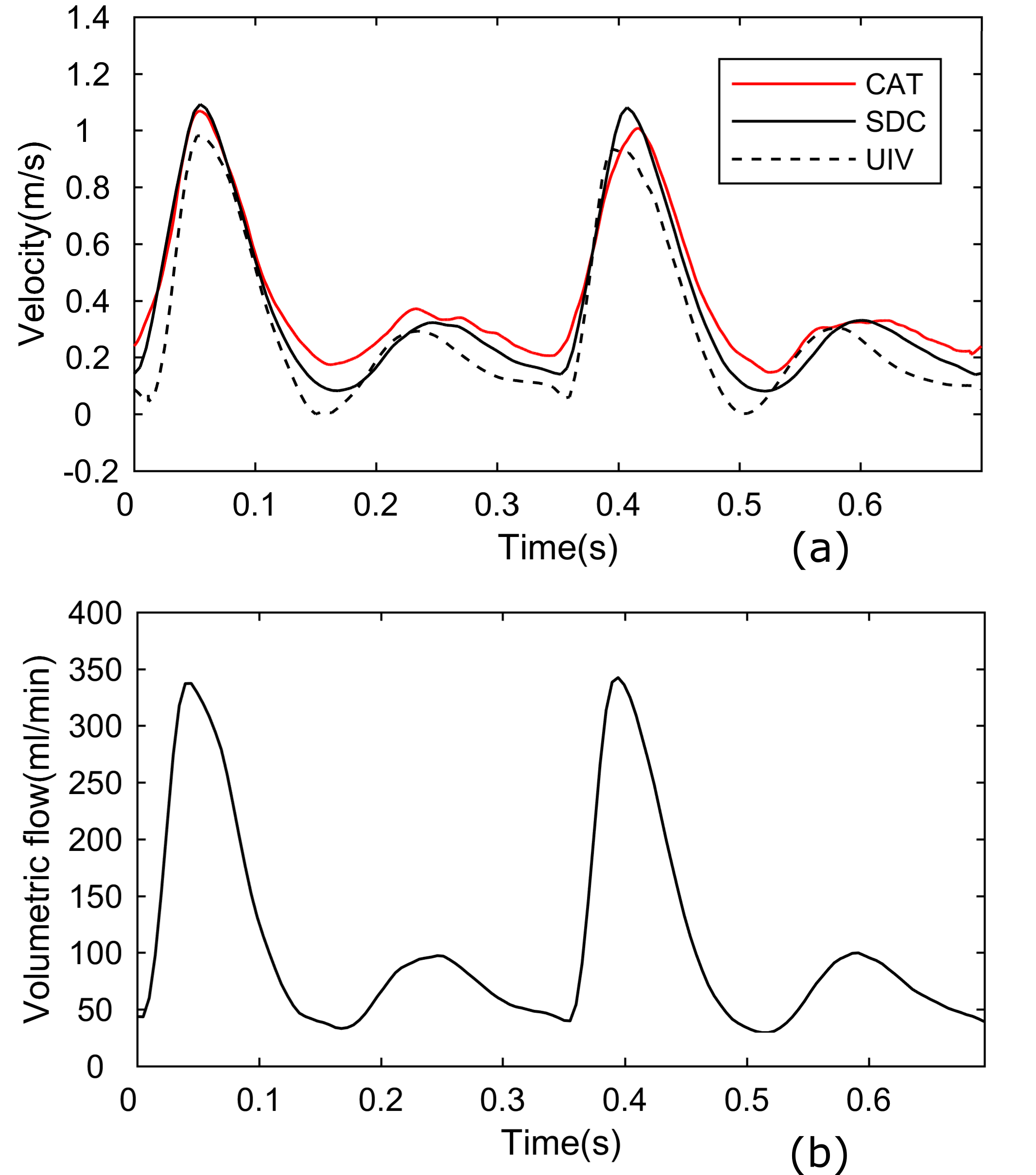
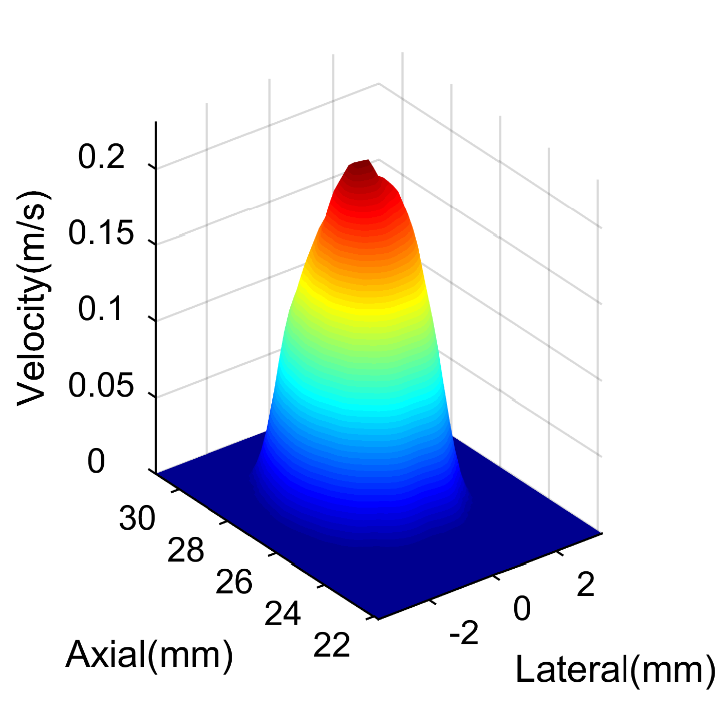
Figure shows measurements of blood velocity in the rabbit aorta (a): SDC curve is the mean velocity within a 1-mm wide square area crossing the center of the aorta in the transverse view. The CAT curve is from the invasive catheter. The UIV curve is the mean velocity within a 1-mm wide square area crossing the center of the aorta in the longitudinal view (b): the volumetric flow rate curve over the aorta calculated from the SDC method. SDC represents the decorrelation method. UIV represents ultrasound imaging velocimetry imaging. CAT represents the catheter.
Supporting Bodies:
This work was supported by the British Heart Foundation under Grant PG/16/95/32350. This work is also partially funded by the UK Engineering and Physical Sciences Research Council under Grant EP/M011933/1.