Ultrasound Motion Artifacts and Correction in High Frame Rate Contrast Imaging
A. Stanziola*, M. Toulemonde*, Y. Li, V. Papadopoulou, R. Corbett, N. Duncan, R J. Eckersley, and M.X. Tang, “MotionArtifacts and Correction in Multi-Pulse High Frame Rate Contrast Enhanced Ultrasound”
IEEE Transactions on Ultrasonics, Ferroelectrics, and FrequencyControl, 2018.
A. Stanziola, M.Toulemonde, Y. Li V. Papadopoulou, and M. X. Tang are with the ULIS Group, Departmentof Bioengineering, Imperial College London, London, SW7 2AZ, UK R. Corbett and N. Duncan are with HammersmithHospital, Imperial College Healthcare NHS Trust London W12 0HS. R. J. Eckersley are with theBiomedical Engineering Department, Division of Imaging Sciences, King’s CollegeLondon, SE1 7EH, London, UK
Abstract:
High frame-rate (HFR) ultrasoundimaging and contrast enhanced ultrasound (CEUS) are often implemented using multi-pulse transmissions, to enhance image quality. Multi-pulse approach,however, suffer from degradation in presence of motion, especially when coherent compounding and CEUS are combined. In this paper, we investigate this effect on the intensity of HFR CEUS in deep tissue imaging using simulations and in vivo contrast echocardiography (CE). Simulation results show that motion artefact is much higher when flow is in axial direction than that lateral direction. Using a pulse repetition frequency suitable for cardiac imaging, amotion of 35 cm/s can cause as much as 28.5dB decrease in image intensity,where compounding can contribute up to 18.7dB of intensity decrease (11angles). This motion effects are also demonstrated for in-vivo cardiac HFR CE, where the large velocities of both the myocardium and the blood are present. Intensity reductions of 10.4 dB are readily visible in the chamber. Finally we demonstrate how performing motion-correction before PI compounding greatlyreduces such motion artefact and improve image signal to noise ratio and contrast.
Supporting Bodies:
This work was supported by an Engineering and Physical Sciences Research Council (EPSRC) grant EP/M011933/1 and grant EP/M010961/1 and by the British Heart Foundation Centre of Research Excellence grant RE/13/4/30184. We gratefully acknowledge the support of NVIDIA Corporation with the donation of the Titan Xp GPU used for this research.
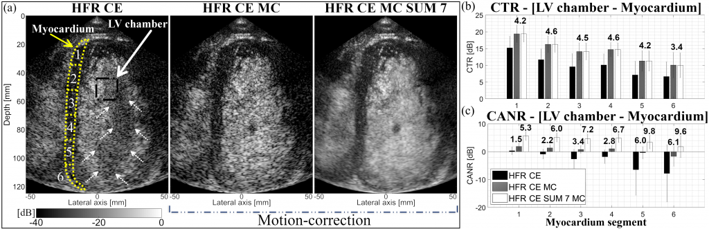
(a) HFR CE images of the left ventricular heart chamber of a human volunteer. The first image shows an HFR CE frame, where dashed arrows indicate the area of motion artefact, the middle one is obtained after motion-correction (MC) and the last one is the result of an average of 7 MC consecutive frames. (b) CTR and (c) CANR corresponding to the different ROIs shown in (a). The bold values are the dB improvement of each technique compared to HFR CE approach.
HFR CE videos of the left ventricular heart chamber of a human volunteer (Top) without and (Bottom) with motion correction. Left images are without temporal average while the right images are created using SUM 7 corresponding to the average of 7 consecutives envelope detected HFR CE frames, with a triangular window centered on the interested frame. All videos are normalized and log compress by their own maximum and display with a dynamic range of 40 dB.