Link: https://ieeexplore.ieee.org/abstract/document/9955557/
Authors: Kai, Riemer, Kai, Matthieu Toulemonde, Jipeng Yan, Marcelo Lerendegui, Elanor Stride, Peter D. Weinberg, Christopher Dunsby, Meng-Xing Tang
Journal: IEEE Transactions on Medical Imaging
doi = 10.1109/TMI.2022.3223554
Abstract
Perfusion by the microcirculation is key to the development, maintenance, and pathology of tissue. Its measurement with high spatiotemporal resolution is consequently valuable but remains a challenge in deep tissue. Ultrasound Localization Microscopy (ULM) provides very high spatiotemporal resolution, but the use of microbubbles requires low contrast agent concentrations, a long acquisition time, and gives little control over the spatial and temporal distribution of the microbubbles. The present study is the first to demonstrate Acoustic Wave Sparsely-Activated Localization Microscopy (AWSALM) and fast-AWSALM for in vivo super-resolution ultrasound imaging, offering contrast on demand and vascular selectivity. Three different formulations of acoustically activatable contrast agents were used. We demonstrate their use with ultrasound mechanical indices well within recommended safety limits to enable fast on-demand sparse activation and destruction at very high agent concentrations. We produce super-localization maps of the rabbit renal vasculature with acquisition times between 5.5 s and 0.25 s, and a 4-fold improvement in spatial resolution. We present the unique selectivity of AWSALM in visualizing specific vascular branches and downstream microvasculature, and we show super-localized kidney structures in systole (0.25 s) and diastole (0.25 s) with fast-AWSALM outdoing microbubble-based ULM. In conclusion, we demonstrate the feasibility of fast and selective measurement of microvascular dynamics in vivo with subwavelength resolution using ultrasound and acoustically activatable nanodroplet contrast agents.
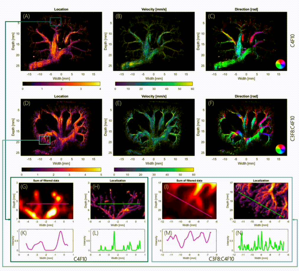
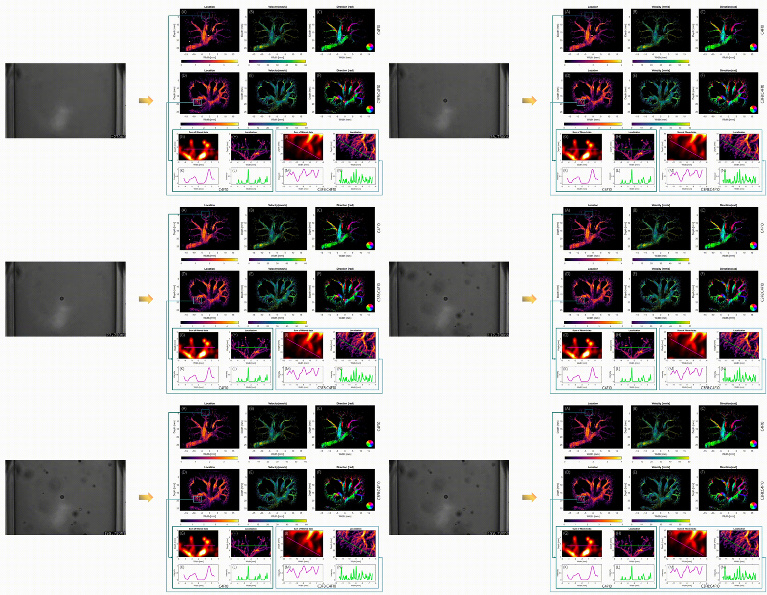
Figure 1 – The left shows nanodroplets inside a cellulose tube under a microscope and their expansion following three acoustic activation pulses. The right side shows AWSALM super-resolution square rooted density map of localization (A,D), absolute flow velocity (B,E) and direction of droplet movement (C,F) in two rabbit kidneys.
Figure 2 – The deliberate activation and deactivation of nanodroplets with AWSALM can highlight different regions of the renal vasculature. Here we focus in three different regions to activate nanodroplets separately from each other.
Figure 3 – The activation of nanodroplets over the full field of view with fast-AWSALM can create sub-second visualizations of the renal vasculature, allowing separation of 0.25 second segments of systole (E) and diastole (F) from a single 1 s long acquisition (D) and outperforms the microbubble comparison (A-C).


