Lluís Guasch, Oscar Calderón Agudo, Meng-Xing Tang, Parashkev Nachev & Michael Warner, “Full-waveform inversion imaging of the human brain”
npj Digital Medicine 3, 28 (2020)
DOI: https://doi.org/10.1038/s41746-020-0240-8
News: https://www.imperial.ac.uk/news/195954/seismic-imaging-technology-could-deliver-detailed/
Affiliation
Lluís Guasch, Oscar Calderón Agudo and Michael Warner are with the Department of Earth Science and Engineering, Imperial College London, United Kingdom.
Meng-Xing Tang is with the Department of Bioengineering, Imperial College London, United Kingdom.
Parashkev Nachev is with the Institute of Neurology, University College London, United Kingdom.
Abstract:
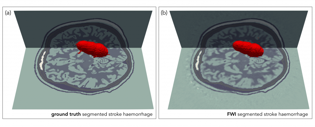
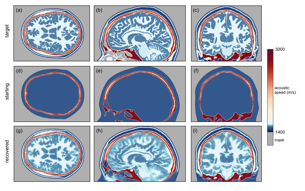
Magnetic resonance imaging and X-ray computed tomography provide the two principal methods available for imaging the brain at high spatial resolution, but these methods are not easily portable and cannot be applied safely to all patients. Ultrasound imaging is portable and universally safe, but existing modalities cannot image usefully inside the adult human skull. We use in silico simulations to demonstrate that full-waveform inversion, a computational technique originally developed in geophysics, is able to generate accurate three-dimensional images of the brain with sub-millimetre resolution. This approach overcomes the familiar problems of conventional ultrasound neuroimaging by using the following: transcranial ultrasound that is not obscured by strong reflections from the skull, low frequencies that are readily transmitted with good signal-to-noise ratio, an accurate wave equation that properly accounts for the physics of wave propagation, and adaptive waveform inversion that is able to create an accurate model of the skull that then compensates properly for wavefront distortion. Laboratory ultrasound data, using ex vivo human skulls and in vivo transcranial signals, demonstrate that our computational experiments mimic the penetration and signal-to-noise ratios expected in clinical applications. This form of non-invasive neuroimaging has the potential for the rapid diagnosis of stroke and head trauma, and for the provision of routine monitoring of a wide range of neurological conditions.
Supporting Bodies:
This work was funded by the Excellence Fund for Frontier Research, a fund created by Imperial College London to initiate and support research in frontier areas that cross traditional disciplinary boundaries. P.N. is funded by the Wellcome Trust (213038/Z/18/Z) and the UCLH NIHR Biomedical Research Centre.